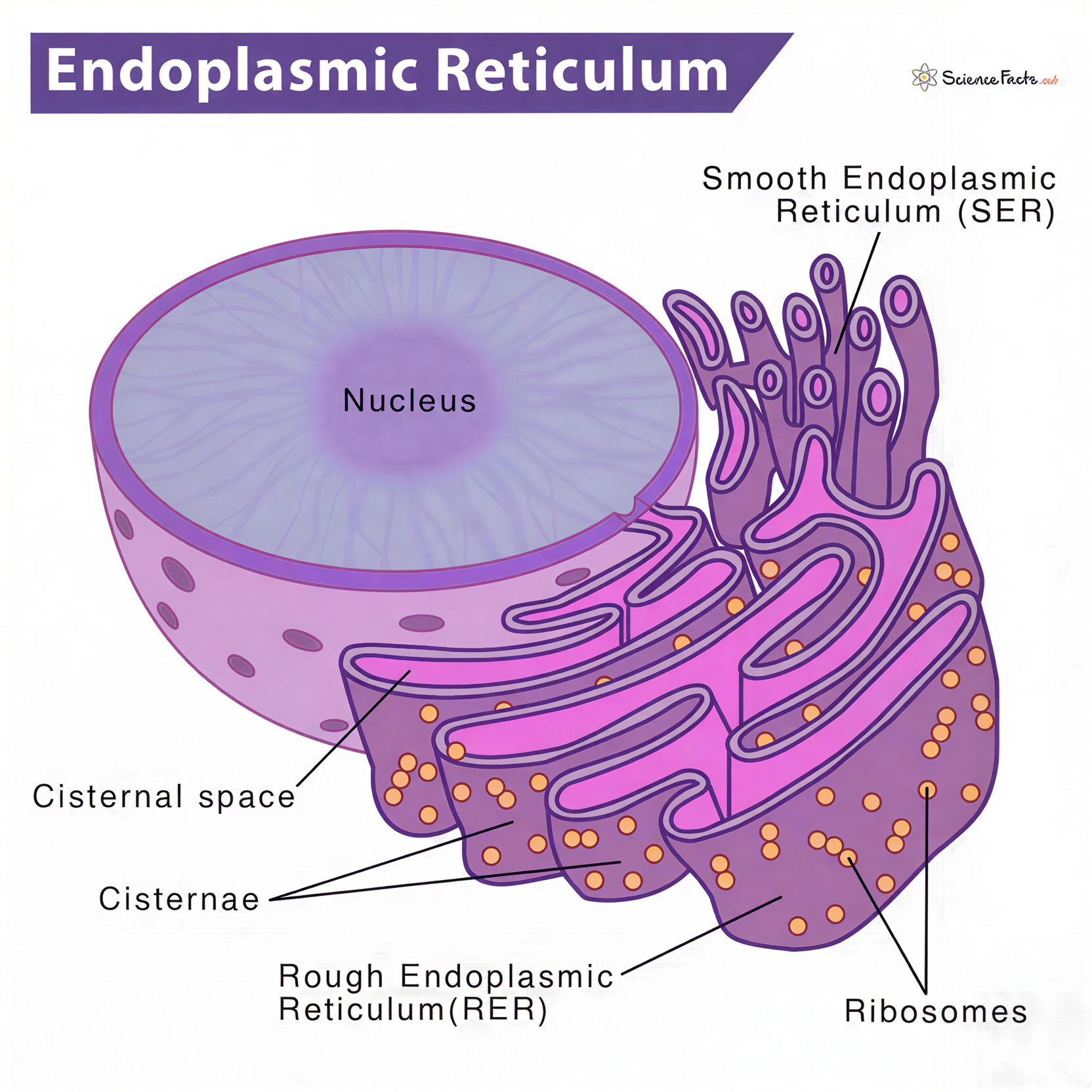
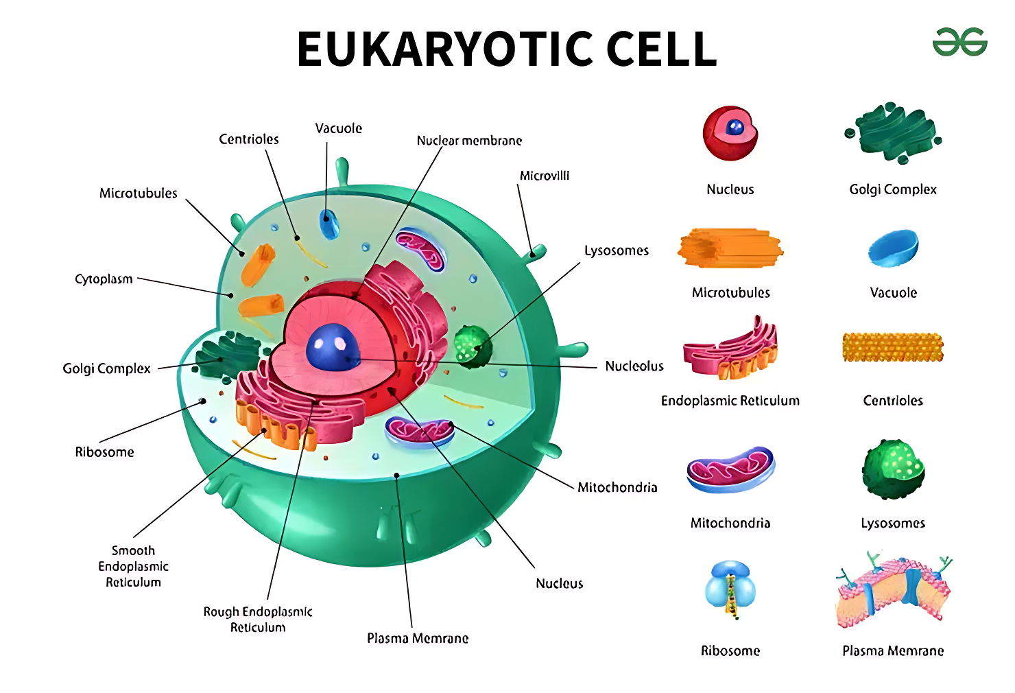
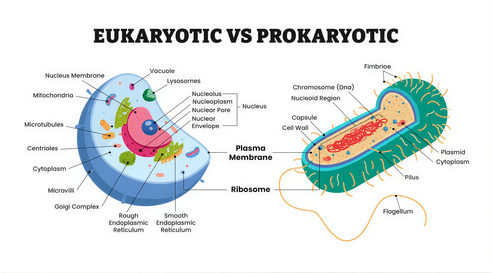
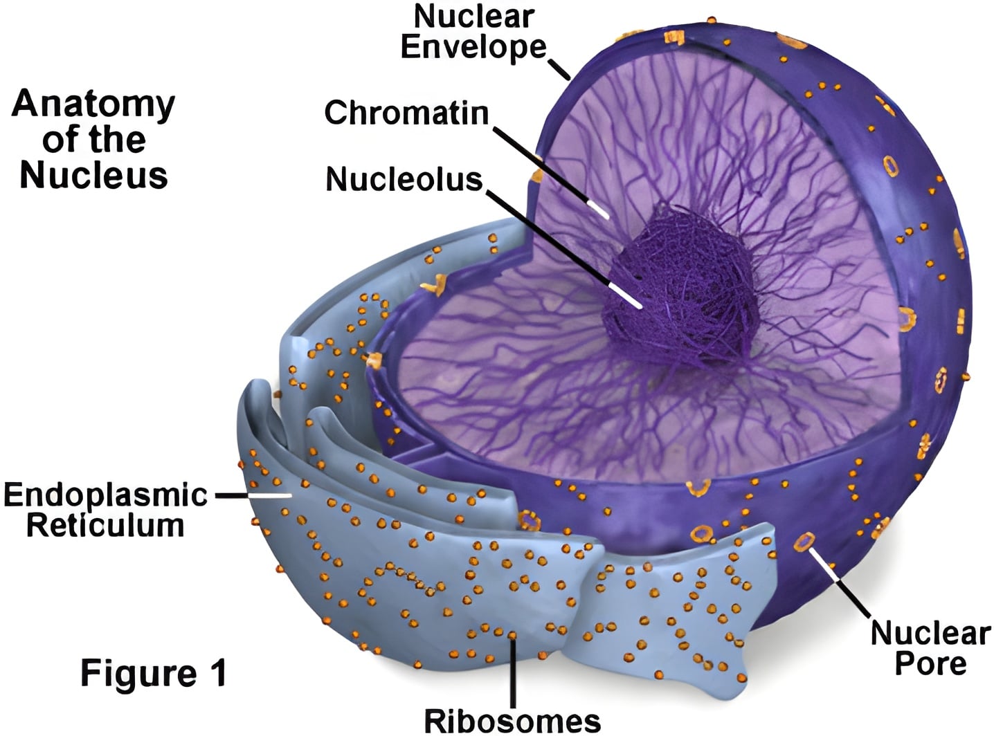
CELL ORGANELLES

Structure • Function • Approximate Size

Nucleus diagram 5–10 µm

Nucleus

The nucleus is a membrane-bound organelle that stores genetic material (DNA) and acts as the control center of the cell.

  • Surrounded by a double membrane called the nuclear envelope
  • Contains nucleolus for rRNA synthesis
  • Absent in prokaryotes and mammalian RBCs
DNA Storage Transcription rRNA
Mitochondria diagram 0.5–1 µm

Mitochondria

Mitochondria are double-membrane organelles responsible for ATP synthesis through oxidative phosphorylation.

  • Inner membrane folded into cristae
  • Contains its own circular DNA
  • Site of aerobic respiration
ATP Respiration DNA
Endoplasmic Reticulum diagram ~0.03–0.05 µm

Endoplasmic Reticulum (ER)

The endoplasmic reticulum is an extensive membranous network involved in synthesis, modification, and transport of biomolecules inside the cell.

  • Rough ER contains ribosomes → protein synthesis
  • Smooth ER lacks ribosomes → lipid & steroid synthesis
  • Forms intracellular transport channels
  • Detoxification of drugs (liver cells)
Protein Synthesis Lipid Metabolism Transport
Golgi apparatus diagram 1–3 µm

Golgi Apparatus

The Golgi apparatus modifies, packages, and sorts proteins and lipids before they are transported to their final destination.

  • Consists of flattened membrane-bound sacs called cisternae
  • Has cis (receiving) and trans (shipping) faces
  • Essential for secretion and membrane renewal
Protein Packaging Vesicles Secretion
Lysosome diagram 0.1–1.2 µm

Lysosome

Lysosomes are enzyme-filled vesicles responsible for intracellular digestion, recycling worn-out organelles and destroying pathogens.

  • Contain hydrolytic enzymes active at acidic pH
  • Formed from Golgi-derived vesicles
  • Referred to as the “suicidal bags” of the cell
Digestion Autophagy Defense
Ribosome diagram 0.02–0.03 µm

Ribosome

Ribosomes are non-membranous particles responsible for protein synthesis.

  • Composed of rRNA and proteins
  • Found free in cytosol or attached to RER
  • Present in both prokaryotic and eukaryotic cells
Protein Synthesis rRNA Translation
Vacuole diagram 5–90 µm

Vacuole

The vacuole is a large membrane-bound sac that maintains turgor pressure, stores substances, and regulates the internal environment of plant cells.

  • Bounded by a selectively permeable membrane called tonoplast
  • Maintains osmotic balance and cell rigidity
  • Plays role in storage and detoxification
Storage Turgor Osmoregulation
Chloroplast diagram 4–10 µm

Chloroplast

Chloroplasts are double-membrane organelles responsible for photosynthesis, converting light energy into chemical energy.

  • Contains chlorophyll pigments
  • Has thylakoids arranged into grana
  • Possesses its own DNA and ribosomes
Photosynthesis Chlorophyll Plastid
Centrosome diagram ~0.5 µm

Centrosome

The centrosome acts as the microtubule-organizing center of the cell and plays a crucial role during cell division.

  • Composed of a pair of perpendicular centrioles
  • Forms spindle fibers during mitosis and meiosis
  • Absent in most higher plant cells
Cell Division Spindle Fibers Microtubules
Cytoskeleton diagram ~0.007-0.025 µm

Cytoskeleton

The cytoskeleton is a dynamic network of protein filaments that maintains cell shape, enables movement, and organizes internal components.

  • Made of microtubules, microfilaments, and intermediate filaments
  • Helps in intracellular transport
  • Essential for cell division and motility
Cell Shape Movement Transport
Plasma membrane diagram ~0.007–0.01 µm

Plasma Membrane

The plasma membrane is a selectively permeable lipid bilayer that encloses the cell and regulates movement of substances.

  • Composed of phospholipid bilayer with proteins
  • Maintains cell integrity and shape
  • Allows selective transport and cell signaling
Selective Permeability Fluid Mosaic Cell Boundary
Cell wall diagram ~0.1–0.5 µm

Cell Wall

The cell wall is a rigid outer covering found outside the plasma membrane that provides protection, shape, and mechanical strength.

  • Made of cellulose in plants
  • Fully permeable to most substances
  • Prevents osmotic bursting of the cell
Cellulose Protection Structural Support
Prokaryotic cell diagram 0.5-5 µm

Prokaryotic Cell

Prokaryotic cells lack a true nucleus and membrane-bound organelles. Genetic material lies freely in the cytoplasm.

  • No nuclear membrane (nucleoid present)
  • Cell wall made of peptidoglycan
  • Ribosomes are 70S
Bacteria No Nucleus 70S Ribosome
Eukaryotic cell diagram 10–100 µm

Eukaryotic Cell

Eukaryotic cells possess a true nucleus and membrane-bound organelles, allowing compartmentalization of cellular functions.

  • True nucleus with nuclear envelope
  • Membrane-bound organelles present
  • Ribosomes are 80S
Nucleus 80S Ribosome Complex Cell
Prokaryotic vs Eukaryotic Cell diagram

Prokaryotic v/s Eukaryotic

A visual comparison highlighting the fundamental structural differences between prokaryotic and eukaryotic cells.